2023.03.13
Spontaneous Activity in Whisker-Innervating Region of Neonatal Mouse Trigeminal Ganglion
研究論文助成事業 採択年度: 2022
Piu Banerjee
Spontaneous Activity in Whisker-Innervating Region of Neonatal Mouse Trigeminal Ganglion
掲載誌:Scientific Reports 発行年: 2022
DOI: 10.1038/s41598-022-20068-z

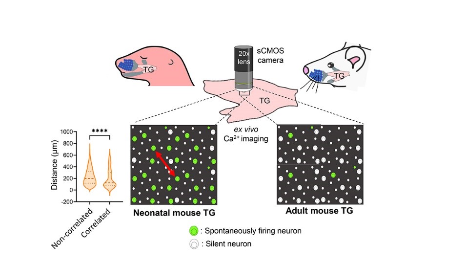
Sensory neurons of neonatal mouse (left) trigeminal ganglion (TG) exhibit spontaneous activity (represented by the green cells) ex vivo, which majorly diminishes by adult stage (right). The entire TG organ is isolated and imaged under a sCMOS camera using a 20x water immersion lens in the established ex vivo system. Violin plot: Correlated neurons are closer to one-another, compared to non-correlated ones (Red arrow: Distance between two spontaneously active neurons).
Spontaneous activity is the activity that occurs without any external sensory input. During the early postnatal period in mammals, such activity is thought to be crucial for the establishment of mature neural circuits. It remains unclear if the peripheral structure of the developing somatosensory system exhibits spontaneous activity, similar to that observed in the retina and cochlea of developing visual and auditory systems, respectively. By establishing an ex vivo calcium imaging system, here I discovered that neurons in the whisker-innervating region of the trigeminal ganglion (TG) of neonatal mice generate spontaneous activity, as indicated by the green-coloured neurons in the graphical abstract. The violin plot demonstrates that a small percentage of neurons showed some obvious correlated activity, and these neurons were mostly located close to one another[C1] [PB2] (Dashed line: median, dotted lines: quartiles, ****: p value<0.0001). TG spontaneous activity was majorly exhibited by medium-to-large diameter neurons, a characteristic of mechanosensory neurons, and was blocked by removal of extracellular calcium. Moreover, this activity was diminished by the adult stage. Spontaneous activity in the TG during the first postnatal week could be a source of spontaneous activity observed in the neonatal mouse barrel cortex.
[C1]At this point, I would suggest you to use the image above to illustrate your discovery. For instance, you can write, “As shown in red arrow in the figure, we found xxxxxxx.”
[PB2]Resolved
書誌情報
- タイトル:Spontaneous Activity in Whisker-Innervating Region of Neonatal Mouse Trigeminal Ganglion.
- 著者:Piu Banerjee, Fumi Kubo, Hirofumi Nakaoka, Rieko Ajima, Takuya Sato, Tatsumi Hirata, and Takuji Iwasato.
- 掲載誌:Scientific Reports
- 掲載年:2023
- DOI:10.1038/s41598-022-20068-z
生命科学研究科 遺伝学専攻 Piu Banerjee
I am a final year graduate student from the Laboratory of Mammalian Neural Circuits, Department of Genetics, SOKENDAI.
